
 Japanese
RESEARCH NEWS Scientists at Nagoya Institute of Technology Uncover Molecular Basis of Red-Green Color Vision
Category:News|Publishing : July 22, 2026
Human color vision depends on three types of cone cells in the retina. Although all three contain the same light-absorbing molecule, 11-cis-retinal, differences in the surrounding protein determine sensitivity to red, green, or blue light. Understanding how these subtle protein differences produce such precise color discrimination has been difficult because cone pigments are highly unstable and structurally challenging to study.
Now, researchers led by Associate Professor KATAYAMA Kota from Nagoya Institute of Technology, Japan, have examined red and green cone pigments from the crab-eating macaque (Macaca fascicularis), whose color vision system closely resembles that of humans, and found that just three amino acid substitutions account for nearly the entire 30 nm difference in light absorption between the two pigments.
The research team included OLIVUCCI Massimo from the University of Siena and Bowling Green State University, KATO Hideaki from The University of Tokyo, and KANDORI Hideki from Nagoya Institute of Technology. This study was published in Science in Volume 392, Issue 6805 on June 25, 2026.
"This study reports the first three-dimensional structures of primate red- and green-sensitive cone visual pigments in their dark state. In the long term, this knowledge may help researchers understand how genetic variations alter color perception and contribute to color vision deficiencies and other visual disorders," says Dr. KATAYAMA.
Macaque and human red and green cone pigments share about 98% of their amino acid sequence and retain key residues known to influence color sensitivity, making them useful models for studying human color vision.
To determine why red and green cone pigments absorb different wavelengths of light, the researchers compared their structures and built detailed computer models of the region of the protein that holds the light-absorbing retinal chromophore. They focused on three amino acid positions that differ between the pigments, created mutant proteins in which these residues were replaced with those found in the red pigment, and analyzed how the substitutions affected light absorption.
The experiments revealed that the red-green spectral shift is primarily governed by three amino acid substitutions--A180S, F277Y, and A285T. These substitutions do not substantially change the shape of the retinal chromophore. Instead, they alter the electrostatic environment surrounding it, causing each pigment to absorb slightly different wavelengths of light. The largest effect comes from placing a threonine residue with a hydroxyl group near the retinal chromophore.
The researchers also discovered a previously unknown membrane-facing opening in cone pigments that is absent in rhodopsin, the rod-cell pigment responsible for low-light vision. This opening may help explain why cone pigments regenerate much faster, enabling continuous color vision in bright light.
"Small sequence changes tune color by reshaping the electrostatic environment around the retinal chromophore, whereas distinct membrane-access pathways enable efficient retinal exchange and rapid visual pigment regeneration," said Dr. KATAYAMA.
These findings show how subtle changes in the chromophore's surroundings and the structure of cone pigments enable both precise color discrimination and rapid regeneration. According to the researchers, these structural insights may also inform future drug discovery efforts targeting visual photoreceptors and other G protein-coupled receptors.
Reference
|
Title of original paper |
Structural insights into spectral tuning and retinal exchange in cone visual pigments |
|
Journal |
Science |
|
DOI |
10.1126/science.adz3996 |
|
Latest Article Publication Date |
June 25, 2026 |
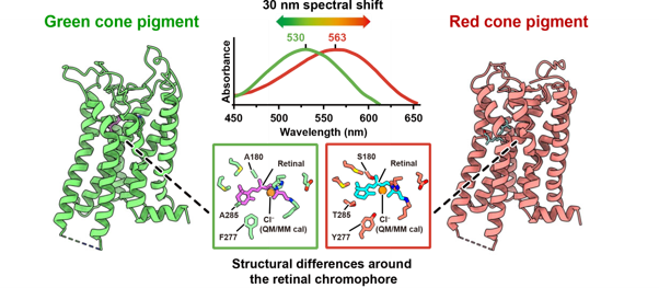
Caption: In macaque cone pigments, just three amino acid substitutions (A180S, F277Y, and A285T) account for nearly the entire 30 nm shift in light absorption underlying red-green color discrimination.
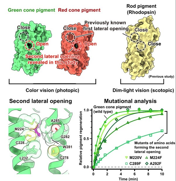
Caption: 3D structures of macaque red and green cone pigments and bovine rhodopsin, revealed by cryo-electron microscopy, show lateral membrane-accessible pores that differ between states, highlighting structural adaptations for rapid color vision in cones and dim-light vision in rods.
About Assistant Professor KATAYAMA Kota from Nagoya Institute of Technology, Japan
Dr.KATAYAMA Kota is an Associate Professor of Life Science and Applied Chemistry at Nagoya Institute of Technology, where he earned his Ph.D. in Engineering in 2013. He leads the KATAYAMA Lab, which focuses on understanding how cone visual pigments enable color vision at the molecular level. Dr. KATAYAMA has published widely in the field and received the 2024 NITech Staff Special Medal. His long-term goal is to uncover the molecular principles of color vision and to provide a structural foundation for future studies of visual disorders and therapeutic development.
Funding information
This work was supported by the Nakayama Foundation for Human Science; the Nakajima Foundation; the Research Foundation for Opto-Science and Technology; the Astellas Foundation for Research on Metabolic Disorders; the Kazato Research Foundation; the Japan Agency for Medical Research and Development (AMED, grant nos. 24bm1123057h0001, 26ak0101309h0001, JP23ama121002, JP24ama121002, JP23ama121001, and JP24ama121001); JSPS KAKENHI (grant nos. 20H05440, 21H04969, 22H00400, 25H01338, 24H02262, 25K09525, 19H03428, 24K02231, 24K18060, 25H02243, 26K18246, 26H00459, 26H00461, 25KJ1446, and 24KJ0981); Grant-in-Aid for Transformative Research Areas (21H05111, 21H05112); MEXT Promotion of Development of a Joint Usage/Research System Project: Coalition of Universities for Research Excellence (CURE, JPMXP1323015482); JST PRESTO (JPMJPR19G4, JPMJPR24OF); JST FOREST (JPMJFR240U, JPMJFR204S); JST ACT-X (JPMJAX222F); and JST CREST (JPMJCR25B5, JPMJCR1753, JPMJCR21P3, JPMJCR23B1). Additional support was provided by the European Union MUR PNRR "National Center for Gene Therapy and Drugs based on RNA Technology" (CN00000041, CN3 RNA - Spoke 6), the US National Science Foundation (CHE-2102619), the Italian Ministry of University and Research (PRIN 2022, 2022K3AY2K), the Center for Photochemical Sciences at Bowling Green State University, the Center for the Evolutionary Origins of Human Behavior (EHUB), Kyoto University, the Cooperative Research Program and Research Units for Exploring Future Horizons at Kyoto University Research Coordination Alliance, and BINDS programs supporting electron microscopy and X-ray crystallography.
Contact
Assistant Professor KATAYAMA Kota
https://katayama-lab.web-ac.jp/
E-mail : katayama.kota[at]nitech.ac.jp
TEL:+8152-735-5218
*Please replace [at] with @ when contacting.